All Images
Before we start
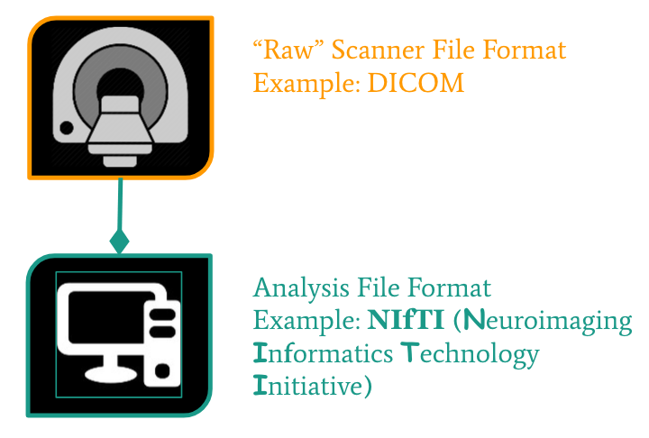
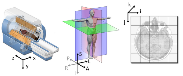
From the scanner to our computer
Figure 1
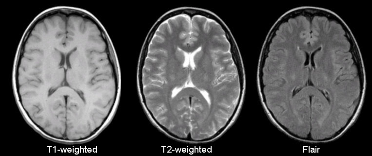
Figure 2
Figure 3
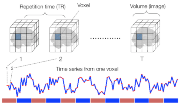
Figure 4

Figure 5
Figure 6
Anatomy of a NIfTI
Figure 1
t1_data contains 3 dimensions. You can think of the data
as a 3D version of a picture (more accurately, a volume). 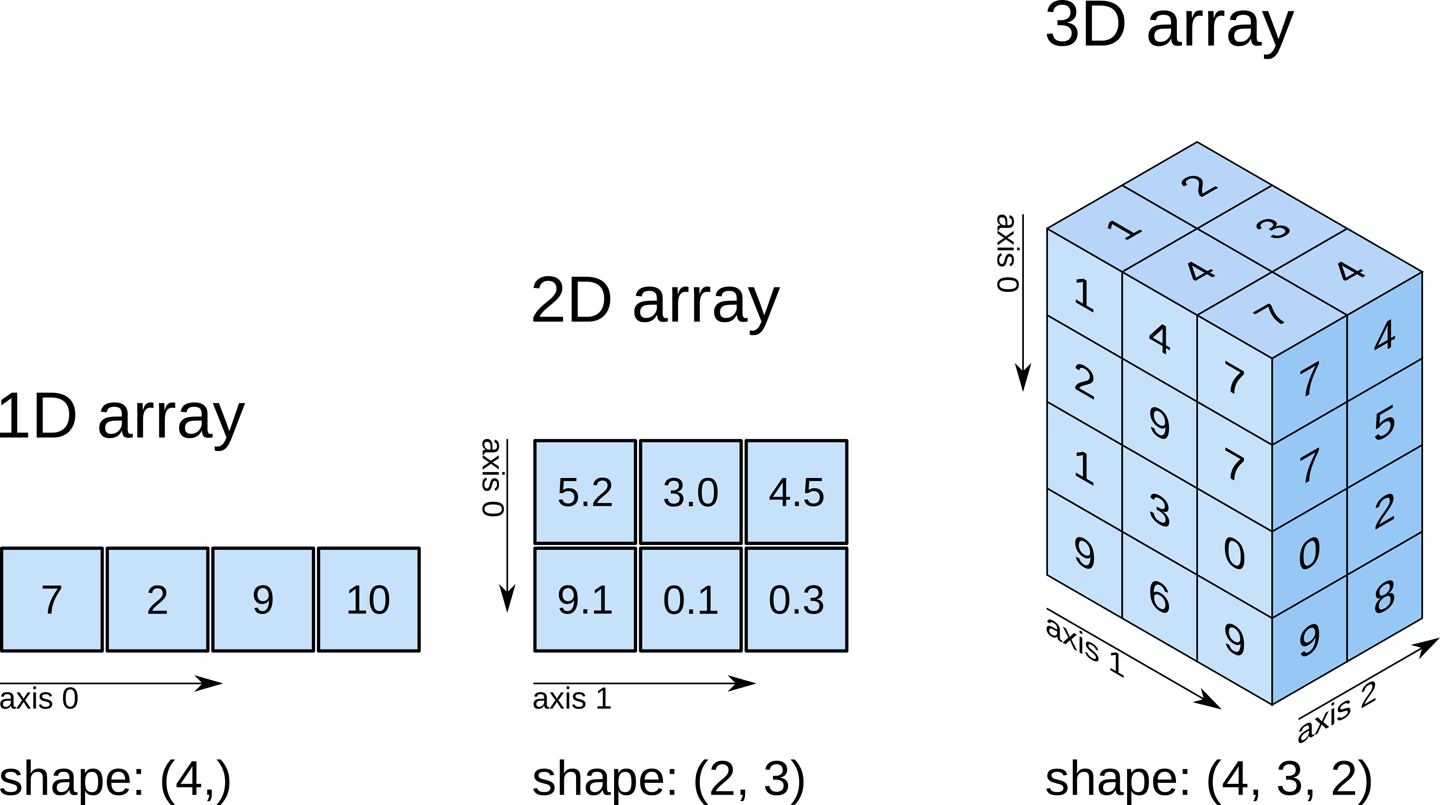
Figure 2
While typical 2D pictures are made out of squares called
pixels, a 3D MR image is made up of 3D cubes called
voxels. 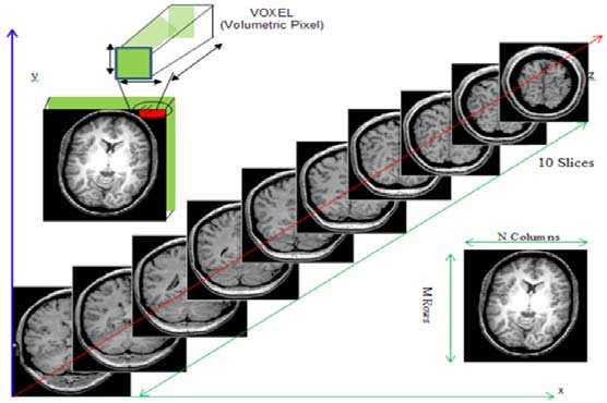
Figure 3
 From left to right:
sagittal, coronal and axial slices.
From left to right:
sagittal, coronal and axial slices.
Figure 4
Data organization with BIDS
Exploring open MRI datasets
BIDS derivatives
Figure 1